Disease journey

Type 2 diabetes

Follow glucose from a meal into the bloodstream, see why insulin’s signal becomes less effective, and connect that core change to tests, medicines, daily care, and complications.

Start with the system

The glucose delivery system loses control

Glucose is fuel carried in the blood. Insulin is not a key that opens every cell; it is a hormone signal that coordinates where fuel is used and stored. In Type 2 diabetes, muscle, liver, and fat tissue respond less effectively to that signal. The pancreas initially compensates by releasing more insulin, but over time its beta cells may no longer supply enough for the body’s needs. Blood glucose then stays elevated.

After a meal

Coordinated response

Pancreas
insulin signal ↓
Muscle

glucose moves into muscle

Liver

liver output slows

Resistance + relative deficiency

Type 2 diabetes

Pancreas
weaker response ↓
Muscle

less muscle uptake

Liver

liver keeps releasing glucose

A schematic, not an anatomical drawing. Insulin acts across many tissues; muscle, liver, fat, pancreas, gut, kidney, and brain all contribute to the full disease.

Coordinated response

  1. 01Carbohydrate is digested and glucose enters the bloodstream.
  2. 02Rising glucose and gut hormones prompt pancreatic beta cells to release insulin.
  3. 03Insulin promotes glucose uptake in muscle and fat and restrains glucose release by the liver.
  4. 04Blood glucose settles toward its usual range as fuel is used or stored.

Type 2 diabetes

  1. 01Muscle, liver, and fat tissue become less responsive to insulin.
  2. 02The pancreas compensates by producing more insulin.
  3. 03The liver continues releasing too much glucose while muscle uptake is reduced.
  4. 04As beta-cell capacity declines, insulin becomes insufficient for the body’s needs and glucose remains elevated.

Core sources: [ada-diagnosis-2026], [niddk-causes], [niddk-resistance]

Reading depth

Chapter 01

Anatomy & physiology

The normal glucose system

Start with the healthy system: where glucose comes from, how the pancreas senses it, and how organs coordinate fuel use between meals.

2 topics · 2 concepts

Study section 1

From meal to cell

Glucose moves through a controlled supply chain involving the intestine, blood, pancreas, liver, muscle, and fat.

Concept 01

What happens after a meal

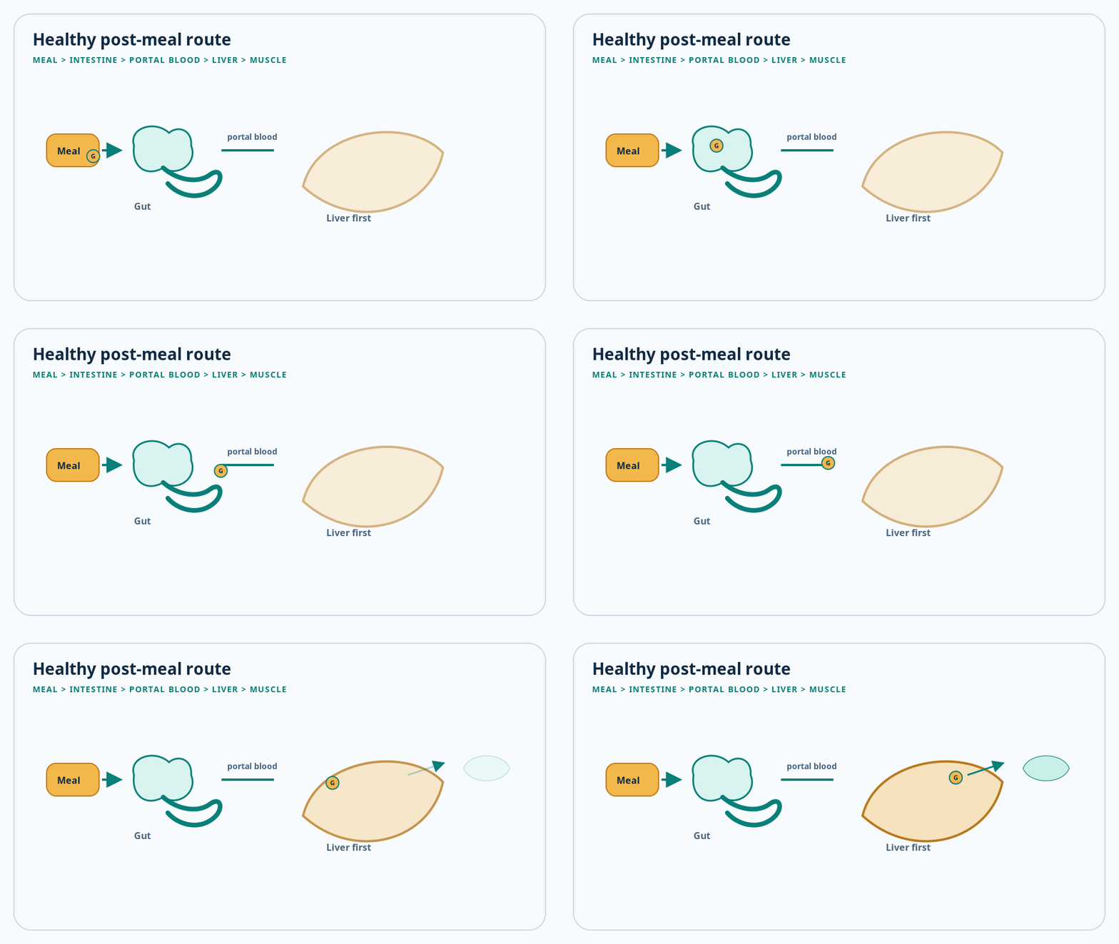
Animated schematic: meal to gut to portal blood to liver — the healthy post-meal glucose route
After a meal: glucose travels intestine → portal blood → liver first

Start here

Digestible carbohydrate is broken into smaller sugars, including glucose, which is absorbed through the small intestine. The resulting rise in blood glucose is a signal—not automatically a problem. It tells the pancreas and other organs that fuel has arrived and needs to be distributed.

Mechanism
  1. 1

    Intestinal enzymes break digestible carbohydrates into absorbable sugars.

  2. 2

    Glucose crosses the intestinal lining and enters portal blood traveling first to the liver.

  3. 3

    The pancreas senses the rise, while gut-derived incretin hormones amplify meal-related insulin release.

  4. 4

    The liver stores some glucose as glycogen; muscle uses or stores glucose according to energy demand.

Clinical connection

A glucose measurement has meaning only in context: fasting, after a glucose load, after a usual meal, or as a longer-term HbA1c estimate. These tests answer different questions.

Hold onto this

  • A post-meal glucose rise is a normal signal that should be regulated.
  • The liver is both a receiver and a producer of glucose.
  • The pancreas responds to nutrients and gut signals, not glucose alone.

Quick check

Which organ receives absorbed glucose first through portal blood?
  1. The liver
  2. The kidney
  3. The thyroid
Reveal answer

The liver. Nutrients absorbed from the intestine travel through the portal circulation to the liver before entering the wider circulation.

Study section 2

The pancreatic control signal

Insulin and glucagon help coordinate whether the body stores fuel or makes it available.

Concept 02

Insulin coordinates several organs

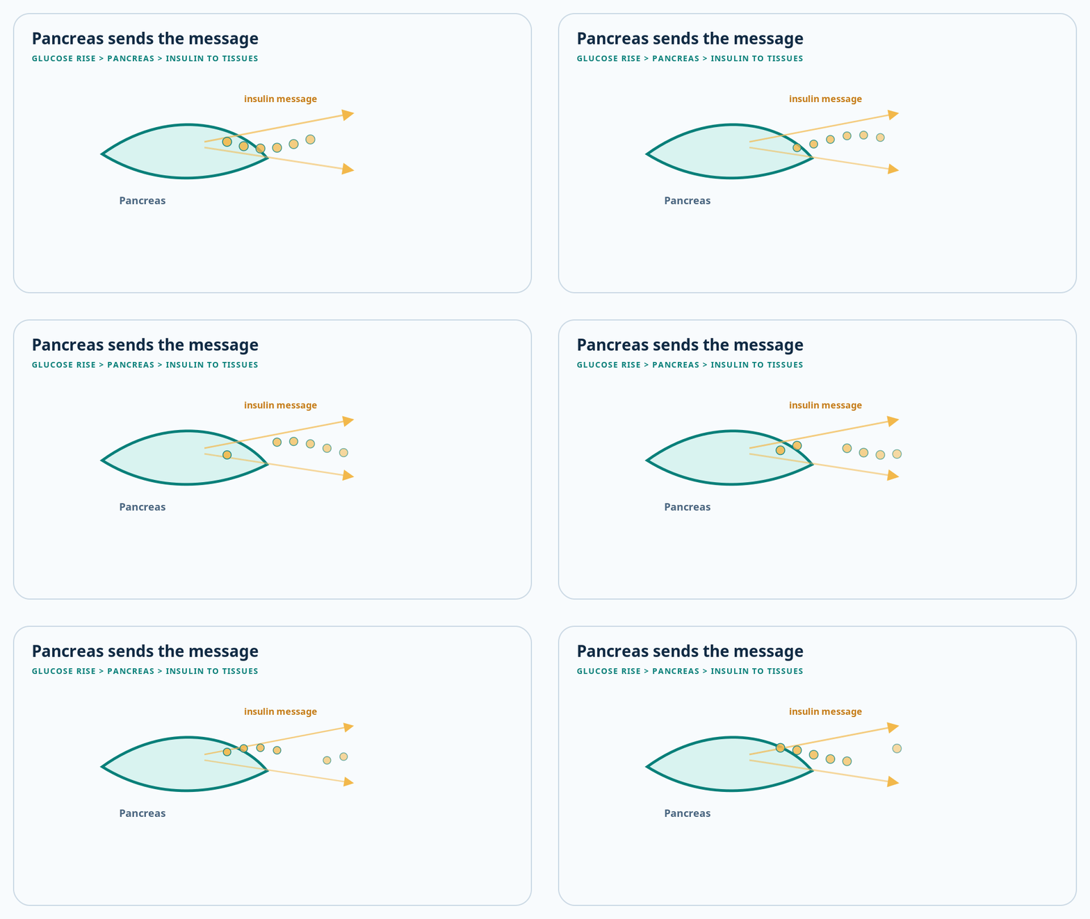
Animated pancreas schematic with an insulin coordinating signal
Pancreas senses the rise and sends an insulin coordinating signal

Start here

Insulin is made by beta cells in pancreatic islets. It signals that fuel is available. In muscle and fat, it supports glucose uptake; in the liver, it helps suppress new glucose output and promotes storage. Glucagon, released by alpha cells, has an opposing role during fasting by supporting hepatic glucose production.

Mechanism
  1. 1

    Beta cells release insulin in response to rising nutrients and incretin signals.

  2. 2

    Insulin promotes GLUT4 movement to the surface of skeletal muscle and adipose cells.

  3. 3

    In the liver, insulin favors glycogen synthesis and restrains gluconeogenesis and glycogen breakdown.

  4. 4

    Between meals, lower insulin and relatively higher glucagon help the liver maintain circulating glucose.

Clinical connection

Type 2 diabetes is not simply the absence of insulin. Many people initially have high insulin levels because the pancreas is compensating for resistance.

Hold onto this

  • Insulin changes metabolism across multiple tissues.
  • Glucagon helps protect against low glucose during fasting.
  • Early Type 2 diabetes may involve excess insulin alongside reduced insulin effect.

Quick check

Why can insulin levels be high early in Type 2 diabetes?
  1. The pancreas is compensating for insulin resistance
  2. The intestine produces insulin
  3. The kidneys stop filtering insulin
Reveal answer

The pancreas is compensating for insulin resistance. Beta cells can increase insulin secretion for a time to overcome reduced tissue responsiveness.

Chapter 02

Biochemistry & cell biology

The signal inside the cell

Zoom in from whole organs to receptors, signaling pathways, transporters, and gut hormones.

2 topics · 2 concepts

Study section 1

From receptor to GLUT4

Insulin binding starts a molecular relay that changes glucose transport and metabolism.

Concept 01

Insulin is a message, not a doorway

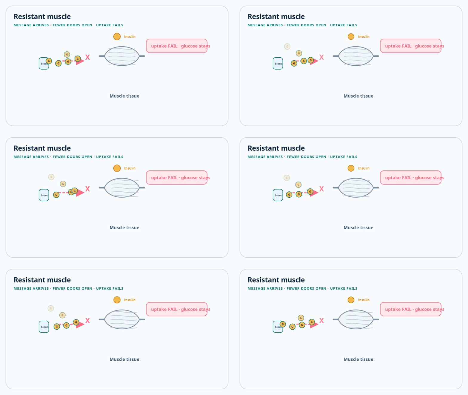
Animated skeletal muscle tissue showing glucose uptake under insulin resistance
Muscle tissue can use or store glucose when insulin signals

Start here

Insulin binds to a receptor on the cell surface. The receptor passes the message through a chain of proteins inside the cell. In muscle and fat, one result is movement of GLUT4 transporters to the cell membrane, where they can carry glucose into the cell.

Mechanism
  1. 1

    Insulin binds the insulin receptor, a receptor tyrosine kinase.

  2. 2

    Receptor activation recruits insulin-receptor-substrate proteins.

  3. 3

    The PI3K–AKT signaling pathway transmits much of the metabolic signal.

  4. 4

    GLUT4-containing vesicles move to the membrane in muscle and adipose tissue, increasing glucose transport.

Clinical connection

Insulin resistance can arise from disruption at several levels and differs between tissues. It is not one broken molecular switch shared identically by every person.

Hold onto this

  • Insulin acts through a signaling cascade.
  • GLUT4 is especially important in skeletal muscle and adipose tissue.
  • Tissue-specific resistance helps explain the varied metabolic picture.
Study section 2

The gut–pancreas conversation

Gut hormones make oral nutrients produce a stronger insulin response than glucose delivered directly into blood.

Concept 02

GLP-1 and GIP announce the meal

3D gut representing meal-related incretin signaling to the pancreas
Gut hormones amplify meal-related insulin release

Start here

After nutrients enter the gut, incretin hormones—especially GLP-1 and GIP—help beta cells produce a glucose-dependent insulin response. Their biology is one reason several modern diabetes medicines target incretin pathways.

Mechanism
  1. 1

    Intestinal cells release GLP-1 and GIP in response to nutrients.

  2. 2

    Both can amplify insulin secretion when glucose is elevated.

  3. 3

    GLP-1 also suppresses inappropriate glucagon, slows gastric emptying, and influences satiety.

  4. 4

    DPP-4 rapidly breaks down native incretin hormones.

Clinical connection

DPP-4 inhibitors slow incretin breakdown, while GLP-1 receptor agonists mimic stronger, longer signaling. These classes are related but are not interchangeable.

Hold onto this

  • The gut participates actively in glucose regulation.
  • Incretin-driven insulin release depends on glucose level.
  • Different drug classes modify this system in different ways.
Chapter 03

Pathology & pathophysiology

How Type 2 diabetes develops

Connect insulin resistance, organ cross-talk, and beta-cell dysfunction into a progression rather than a single event.

2 topics · 2 concepts

Study section 1

Resistance across tissues

Muscle, liver, and fat contribute differently to rising glucose and altered metabolism.

Concept 01

Muscle takes less, liver releases more

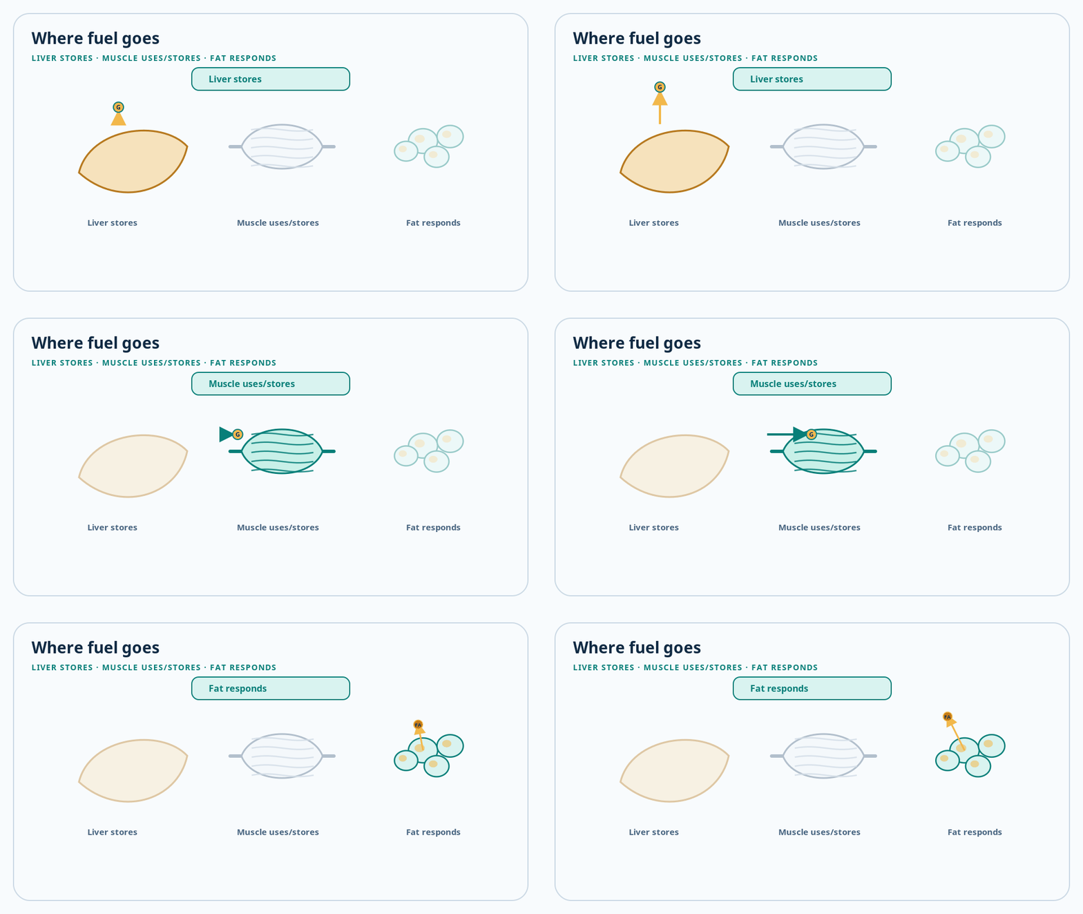
Liver, muscle, and fat schematic showing where fuel goes after a meal
Where fuel goes: liver stores, muscle uses or stores, fat responds

Start here

Insulin resistance means a usual insulin signal produces a smaller biological response. Resistant muscle removes less glucose after meals. The resistant liver fails to fully switch off glucose production. Resistant fat tissue releases more fatty acids, which can further disturb liver, muscle, and beta-cell function.

Mechanism
  1. 1

    Reduced muscle insulin signaling lowers meal-related glucose disposal.

  2. 2

    Hepatic insulin resistance permits continued gluconeogenesis and glycogen breakdown.

  3. 3

    Adipose insulin resistance increases lipolysis and circulating fatty acids.

  4. 4

    Ectopic fat, inflammation, genetics, sleep, medicines, aging, and other factors can influence this network.

Clinical connection

Body size alone cannot diagnose insulin resistance or Type 2 diabetes. Risk reflects genetics, fat distribution, age, environment, medicines, pregnancy history, and other factors.

Hold onto this

  • Insulin resistance is tissue-specific.
  • Fasting glucose is strongly influenced by hepatic glucose output.
  • Type 2 diabetes is multifactorial and should not be reduced to personal blame.

Quick check

Which change most directly contributes to elevated fasting glucose?
  1. The liver continues producing glucose
  2. The intestine stops absorbing all glucose
  3. Muscle releases insulin
Reveal answer

The liver continues producing glucose. Inadequately suppressed hepatic glucose production is a major contributor to fasting hyperglycemia.

Study section 2

Compensation and beta-cell failure

The pancreas can compensate for resistance, but that compensation may not remain sufficient.

Concept 02

Relative insulin deficiency emerges over time

Animated pancreas schematic with an insulin coordinating signal
Pancreas senses the rise and sends an insulin coordinating signal

Start here

At first, beta cells may release extra insulin and keep glucose near the usual range. As resistance increases or beta-cell function declines, compensation becomes incomplete. Glucose may pass through a prediabetes range and later meet diabetes criteria. The pace and pattern vary substantially between people.

Mechanism
  1. 1

    Insulin resistance raises the insulin demand required for glucose control.

  2. 2

    Beta cells initially compensate through increased secretion.

  3. 3

    Genetic susceptibility, glucotoxicity, lipotoxicity, islet stress, and other mechanisms can reduce beta-cell function.

  4. 4

    Relative insulin deficiency becomes more important as the disease progresses.

Clinical connection

Type 2 diabetes has no universally accepted numbered stages. Prediabetes, diagnosed diabetes, complications, and remission are clinical states—not a guaranteed one-way sequence.

Hold onto this

  • Insulin resistance alone does not always produce diabetes.
  • Beta-cell capacity determines whether compensation succeeds.
  • Progression can be slowed, and some people achieve remission, but follow-up remains important.
Chapter 04

Clinical medicine & laboratory science

How clinicians identify it

Learn what each glucose test measures, why confirmation matters, and when another diabetes type should be considered.

2 topics · 2 concepts

Study section 1

Four routes to diagnosis

HbA1c, fasting glucose, an oral glucose tolerance test, and symptomatic random glucose provide different windows on dysglycemia.

Concept 01

A result needs context and confirmation

Animated schematic: meal to gut to portal blood to liver — the healthy post-meal glucose route
After a meal: glucose travels intestine → portal blood → liver first

Start here

Diabetes can be diagnosed using HbA1c or plasma glucose criteria. Standard thresholds include HbA1c at least 6.5%, fasting plasma glucose at least 126 mg/dL, or 2-hour plasma glucose at least 200 mg/dL during a 75-g oral glucose tolerance test. Random plasma glucose at least 200 mg/dL is diagnostic when classic hyperglycemic symptoms or a hyperglycemic crisis are present.

Mechanism
  1. 1

    Fasting glucose samples hepatic glucose regulation after an overnight fast.

  2. 2

    The oral glucose tolerance test stresses the system and measures the 2-hour response.

  3. 3

    HbA1c estimates longer-term glycemic exposure through glucose attachment to hemoglobin.

  4. 4

    Without unequivocal hyperglycemia, diagnosis generally requires confirmatory abnormal testing.

Clinical connection

HbA1c can be misleading when red-cell turnover or hemoglobin is altered. Clinicians choose and interpret tests in light of anemia, hemoglobin variants, pregnancy, kidney disease, medicines, acute illness, and symptoms.

Hold onto this

  • One test does not answer every diagnostic question.
  • Symptoms change how a random glucose result is interpreted.
  • Unexpected or borderline results often require confirmation.

Quick check

In an asymptomatic person without unequivocal hyperglycemia, what is usually needed after one abnormal result?
  1. Confirmatory abnormal testing
  2. Immediate lifelong insulin
  3. No further assessment
Reveal answer

Confirmatory abnormal testing. Confirmation reduces the chance that biological or measurement variation produces a mistaken diagnosis.

Study section 2

Not every adult has Type 2

Diabetes classification is a clinical reasoning task, especially when features are atypical.

Concept 02

Type 1, LADA, monogenic, and secondary diabetes

Start here

Age or body weight alone cannot determine diabetes type. Autoimmune Type 1 diabetes can begin in adults. Monogenic diabetes, pancreatic disease, endocrine disorders, pregnancy, and medicines such as glucocorticoids can produce different forms of diabetes that may need different management.

Mechanism
  1. 1

    Type 1 diabetes involves autoimmune beta-cell destruction and absolute insulin deficiency.

  2. 2

    Slowly progressive autoimmune diabetes in adults may initially resemble Type 2 diabetes.

  3. 3

    Single-gene defects can alter beta-cell function in monogenic diabetes.

  4. 4

    Pancreatic injury, hormonal disease, and medicines can create secondary diabetes.

Clinical connection

Unexpected weight loss, ketosis, rapid deterioration, a strong multigenerational pattern, pancreatic disease, or poor response to the expected treatment can prompt additional classification tests.

Hold onto this

  • Diabetes type is not assigned by appearance.
  • Correct classification changes treatment and family counseling.
  • Atypical features deserve reassessment rather than assumption.
Chapter 05

Pharmacology & therapeutics

How treatments change the system

Map each medicine class back to the organ or signal it changes. Treatment selection is person-centered, not a universal ladder.

2 topics · 3 concepts

Study section 1

Reduce output or improve sensitivity

Several medicines act mainly on hepatic glucose production or tissue responsiveness.

Concept 01

Metformin and thiazolidinediones act differently

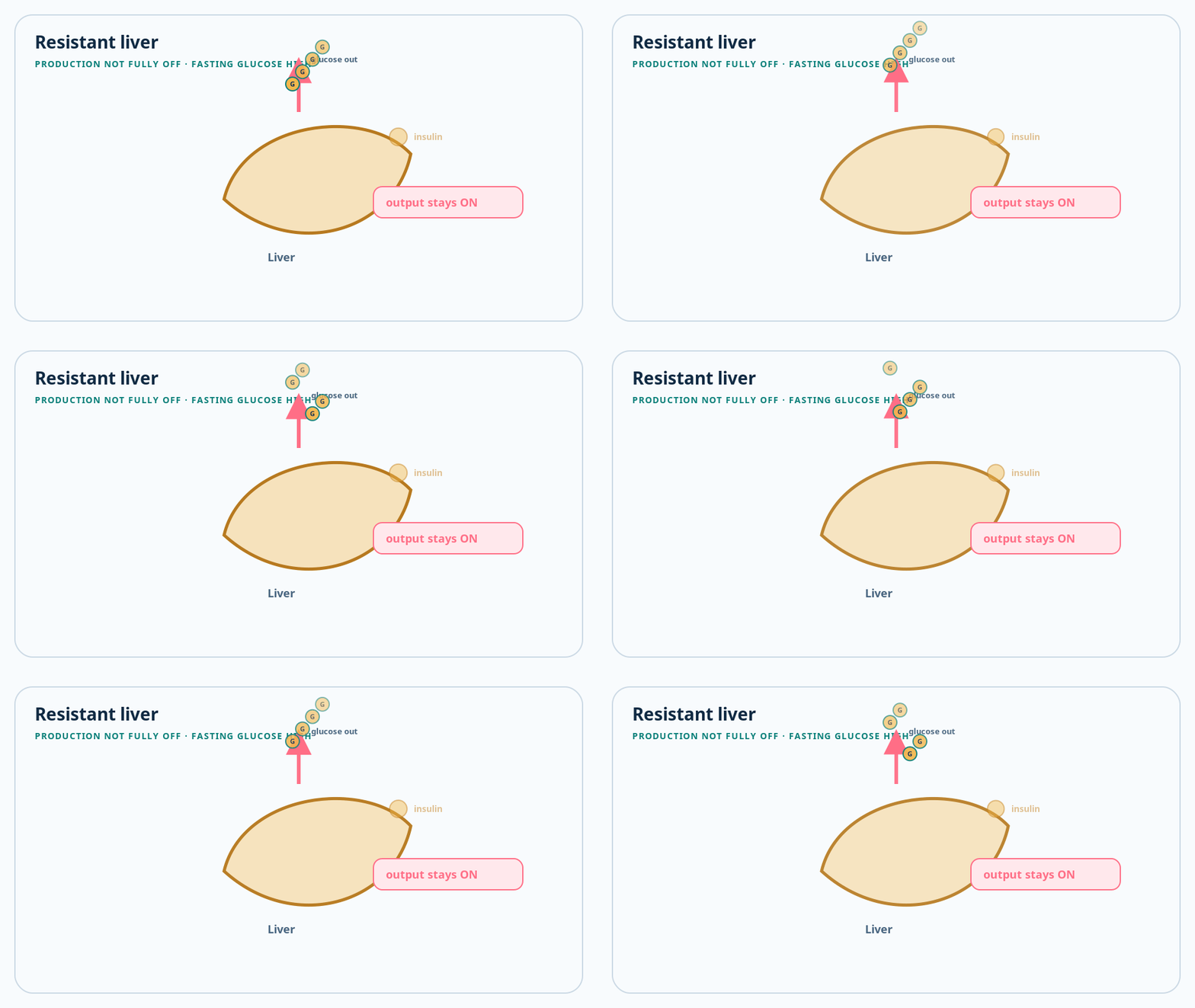
Animated liver schematic showing glucose output that stays on when insulin resistance is present
The liver receives, stores, and can keep releasing glucose

Start here

Metformin primarily lowers excessive hepatic glucose production and can improve insulin sensitivity. Thiazolidinediones activate PPAR-gamma and improve insulin sensitivity in fat, muscle, and liver. Their clinical trade-offs are different even though both address resistance-related biology.

Mechanism
  1. 1

    Metformin reduces hepatic glucose output through several linked cellular mechanisms.

  2. 2

    Thiazolidinediones alter gene transcription through the nuclear receptor PPAR-gamma.

  3. 3

    Neither class directly replaces insulin.

  4. 4

    Kidney, liver, heart, fracture, fluid-retention, tolerability, and cost considerations affect selection.

Clinical connection

Metformin remains widely used and is prominent in Indian workflows, but modern treatment is individualized. Cardiovascular disease, heart failure, kidney disease, weight goals, hypoglycemia risk, cost, and preferences can change the starting choice.

Hold onto this

  • Similar glucose lowering does not mean identical organ effects.
  • Comorbidities matter when selecting a medicine.
  • A clinician must assess contraindications and monitoring needs.
Study section 2

Change insulin release, incretins, or renal glucose handling

Other classes target beta cells, gut hormone pathways, or glucose reabsorption in the kidney.

Concept 02

Four mechanisms, four sets of trade-offs

Start here

Sulfonylureas stimulate insulin release. DPP-4 inhibitors extend native incretin activity. GLP-1 receptor agonists and dual GIP/GLP-1 therapies produce stronger incretin-pathway effects. SGLT2 inhibitors cause the kidney to return less filtered glucose to the blood, so more leaves in urine.

Mechanism
  1. 1

    Sulfonylureas close beta-cell ATP-sensitive potassium channels and can cause glucose-independent insulin release.

  2. 2

    DPP-4 inhibition prolongs endogenous GLP-1 and GIP signaling.

  3. 3

    GLP-1–based therapy enhances glucose-dependent insulin release, suppresses glucagon, influences gastric emptying and satiety, and can reduce weight.

  4. 4

    SGLT2 inhibition reduces proximal-tubule glucose reabsorption and has important heart and kidney benefits in selected people.

Clinical connection

Hypoglycemia and weight gain are important with sulfonylureas; incretin therapies commonly cause gastrointestinal effects; SGLT2 inhibitors can cause genital infections, volume depletion, and rare ketoacidosis. This is why treatment cannot safely be chosen from mechanism alone.

Hold onto this

  • Drug classes target different failure points in the system.
  • Some heart and kidney benefits extend beyond lowering HbA1c.
  • Benefits, harms, access, and personal context must be considered together.

Quick check

Which class lowers glucose by reducing kidney glucose reabsorption?
  1. SGLT2 inhibitors
  2. DPP-4 inhibitors
  3. Thiazolidinediones
Reveal answer

SGLT2 inhibitors. SGLT2 inhibitors act in the proximal renal tubule, increasing urinary glucose excretion.

Concept 03

Insulin replaces an increasingly insufficient signal

Start here

People with Type 2 diabetes may need insulin temporarily or long term. It can be required when hyperglycemia is severe, during acute illness or pregnancy, or when beta-cell secretion no longer meets the body’s needs. Needing insulin is not a personal failure; it reflects physiology and clinical circumstances.

Mechanism
  1. 1

    Injected insulin supplements or replaces endogenous insulin action.

  2. 2

    Basal insulin mainly restrains glucose production between meals and overnight.

  3. 3

    Mealtime insulin addresses nutrient-related glucose rises.

  4. 4

    Insulin plans require individualized dosing, monitoring, injection education, and hypoglycemia prevention.

Clinical connection

Severe symptoms, ketosis, marked hyperglycemia, or acute metabolic decompensation require prompt professional assessment. This lesson intentionally does not provide dosing instructions.

Hold onto this

  • Type 2 diabetes can progress to substantial insulin deficiency.
  • Insulin can be used alongside other medicines.
  • Safe insulin use requires individualized clinical training.
Chapter 06

Preventive medicine & chronic care

Living care and monitoring

Treatment is a repeated feedback loop involving food, activity, sleep, medicines, measurements, risk factors, and access to care.

2 topics · 2 concepts

Study section 1

Lifestyle is treatment, not blame

Daily behaviors can change insulin sensitivity and cardiovascular risk, but recommendations must fit culture, health, resources, and medicines.

Concept 01

Food and movement change the metabolic workload

Liver, muscle, and fat involved when food and movement change glucose workload
Food and movement change the workload on liver, muscle, and fat

Start here

Eating patterns affect how quickly and how much glucose enters circulation. Physical activity increases muscle glucose use and can improve insulin sensitivity. Sleep, tobacco, alcohol, stress, food access, work patterns, and medicines also matter. Sustainable care is designed with the person rather than prescribed as a moral test.

Mechanism
  1. 1

    Active muscle can increase glucose uptake through insulin-dependent and contraction-related pathways.

  2. 2

    Weight reduction, when appropriate and achievable, can lower insulin resistance and may permit remission in some people.

  3. 3

    Diet quality, carbohydrate amount and distribution, fiber, and total energy intake interact rather than acting as one forbidden-food list.

  4. 4

    Blood-pressure, lipid, tobacco, sleep, and psychosocial care reduce risk beyond glucose alone.

Clinical connection

Activity and meal changes can alter hypoglycemia risk when used with insulin or insulin-secretagogue medicines. Individual plans should account for kidney disease, pregnancy, frailty, eating disorders, and other conditions.

Hold onto this

  • Lifestyle intervention is part of medical treatment.
  • There is no single eating plan suitable for everyone.
  • Glucose is only one part of cardiovascular and kidney risk.
Study section 2

Measure, interpret, adjust

Different measurements reveal immediate glucose, longer-term exposure, and organ risk.

Concept 02

Monitoring extends beyond HbA1c

Start here

HbA1c summarizes glycemic exposure but does not show every high, low, or daily pattern. Finger-stick testing or continuous glucose monitoring may answer different questions. Kidney tests, blood pressure, lipids, eye examinations, foot assessment, and medication review look for risks that glucose alone cannot capture.

Mechanism
  1. 1

    HbA1c reflects glycation over the life span of circulating red cells.

  2. 2

    Capillary glucose provides a measurement at one moment.

  3. 3

    Continuous glucose monitoring shows trends and time in ranges.

  4. 4

    Urine albumin and estimated filtration assess different aspects of kidney injury and function.

Clinical connection

Targets are individualized. Age, pregnancy, hypoglycemia risk, duration of diabetes, comorbidities, life expectancy, support, and treatment burden all influence what is appropriate.

Hold onto this

  • No single measurement represents the entire disease.
  • Monitoring should answer a specific clinical question.
  • A target is a shared clinical decision, not a universal score.
Chapter 07

Pathology & internal medicine

Why long-term control matters

Persistent metabolic and vascular stress can affect small vessels, large arteries, nerves, kidneys, eyes, feet, and the brain.

2 topics · 2 concepts

Study section 1

Small vessels and large arteries

Diabetes complications arise through overlapping glucose, blood-pressure, lipid, inflammatory, clotting, and vascular pathways.

Concept 01

One metabolic disease, many organ effects

Liver, muscle, and fat as a multi-organ map for Type 2 diabetes complications
One metabolic disease can stress many organs over time

Start here

Retinopathy, kidney disease, and neuropathy are often grouped as microvascular complications. Heart attack, stroke, and peripheral arterial disease involve larger arteries. These categories overlap: kidney disease increases cardiovascular risk, and nerve and artery disease can combine to threaten the foot.

Mechanism
  1. 1

    Chronic hyperglycemia promotes glycation, oxidative stress, endothelial dysfunction, and altered microvascular signaling.

  2. 2

    Hypertension and abnormal lipids add mechanical and atherosclerotic injury.

  3. 3

    Glomerular injury can cause albumin leakage before filtration falls.

  4. 4

    Sensory loss, altered foot loading, poor perfusion, and infection can combine in diabetic foot disease.

Clinical connection

Screening seeks injury before symptoms appear. Sudden vision change, one-sided weakness, chest pressure, a cold or discolored limb, or a rapidly worsening foot wound needs urgent medical assessment.

Hold onto this

  • Complication prevention is broader than glucose lowering.
  • Kidney and cardiovascular disease are tightly connected.
  • Early screening can find treatable risk before symptoms.
Study section 2

When glucose becomes an emergency

Severe hyperglycemia can cause dehydration and altered consciousness; ketoacidosis can also occur in Type 2 diabetes.

Concept 02

HHS and DKA require emergency care

Start here

Hyperosmolar hyperglycemic state, or HHS, usually involves extreme hyperglycemia, profound dehydration, and changes in alertness with little significant ketoacidosis. Diabetic ketoacidosis, or DKA, involves ketone production and metabolic acidosis. Type 2 diabetes is more commonly associated with HHS, but DKA and mixed states can occur.

Mechanism
  1. 1

    Insufficient effective insulin allows glucose to rise and causes osmotic loss of water and electrolytes in urine.

  2. 2

    Severe dehydration raises blood osmolality and can impair brain function.

  3. 3

    More severe insulin deficiency permits fat breakdown and acidic ketone accumulation.

  4. 4

    Infection, missed insulin, acute illness, myocardial infarction, some medicines, and newly presenting diabetes can precipitate a crisis.

Clinical connection

Confusion, unusual drowsiness, repeated vomiting, deep or rapid breathing, severe weakness, inability to keep fluids down, or signs of major dehydration with high glucose are emergency warning signs. Seek local emergency care rather than relying on an educational app.

Hold onto this

  • HHS and DKA are medical emergencies.
  • Type 2 diabetes does not exclude ketoacidosis.
  • Mental-status change and major dehydration are critical warning signs.

Quick check

Which finding should be treated as an emergency warning sign?
  1. Confusion with severe dehydration
  2. Reading about HbA1c
  3. Feeling hungry before lunch
Reveal answer

Confusion with severe dehydration. Altered mental status with dehydration can signal severe metabolic decompensation and needs emergency assessment.

All sources

Evidence behind this journey

  1. 01Diagnosis and Classification of Diabetes: Standards of Care in Diabetes—2026American Diabetes Association, Diabetes Care · 2026Open source ↗
  2. 02Pharmacologic Approaches to Glycemic Treatment: Standards of Care in Diabetes—2026American Diabetes Association, Diabetes Care · 2026Open source ↗
  3. 03Symptoms & Causes of DiabetesNational Institute of Diabetes and Digestive and Kidney Diseases · 2025Open source ↗
  4. 04Insulin Resistance & PrediabetesNational Institute of Diabetes and Digestive and Kidney Diseases · 2025Open source ↗
  5. 05Diabetes fact sheetWorld Health Organization · 2026Open source ↗
  6. 06Standard Treatment Workflow: Diabetes Mellitus Type 2Indian Council of Medical Research and Department of Health Research · 2024Open source ↗